
UNDERSTANDING YOUR MULTIPLE MYELOMA LAB TESTS
Stay on top of lab work
Your healthcare provider will help you schedule lab tests to track your symptoms and treatment progress. It’s important that you familiarize yourself with the different advanced lab tests and understand what your results mean.

Lab tests you might need
What is an M spike in multiple myeloma?
Multiple myeloma cells make an abnormal antibody called M protein. M protein can accumulate in the blood or urine of people with multiple myeloma and increasing levels can be an early sign of a multiple myeloma relapse.
Blood tests can check for M protein
It can be hard to know if multiple myeloma has returned because it sometimes does not cause any symptoms. Your healthcare team will monitor your multiple myeloma on a regular basis using several different lab tests and imaging. Increasing M protein levels can be identified by an M spike. An M spike is seen on tests when elevated levels of M protein are detected in your blood or urine.
Complete blood count (CBC)
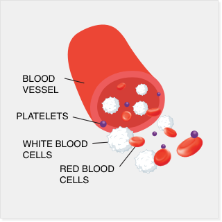
A CBC measures the number of blood cells you have, including red blood cells, white blood cells, and platelets. Myeloma plasma cells can cause low blood counts because they overgrow and crowd out normal cells in the bone marrow. Monitoring blood cell counts can be used to track disease progression and treatment effects.

Blood chemistry profile
A blood chemistry profile measures the levels of different substances in your blood. These levels provide insight to the function of different organs (kidney, liver, etc) that can be affected by multiple myeloma and treatments.
Serum protein electrophoresis (SPEP)
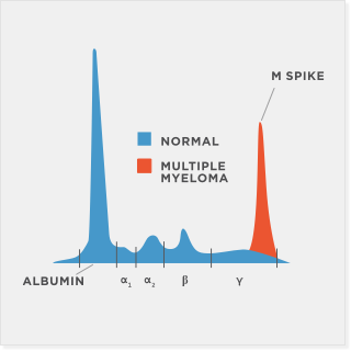
Antibodies are part of your immune system and help your body fight infection. They are produced by healthy plasma cells. In multiple myeloma, one plasma cell becomes cancerous and makes multiple copies of itself (clones). Each copy makes an abnormal antibody called M protein. A test called SPEP is used to separate and identify the presence of M protein in the blood. When M protein is detected, it is called an M spike.
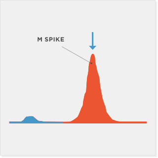
The UPEP test estimates the quantity of certain proteins in your urine. The arrow in this UPEP diagram corresponds to the M spike (M protein). The normal UPEP range is ≤500 mg/24 hours for asymptomatic patients.
Urine protein electrophoresis (UPEP)
The UPEP test estimates the quantity of certain proteins in your urine. The arrow in this UPEP diagram corresponds to the M spike (M protein). The normal UPEP range is ≤500 mg/24 hours for asymptomatic patients.
M spike (myeloma gamma globulin) lab test
The M spike lab test determines the amount of M protein in your blood. The presence of any M spike is abnormal because it reflects the presence of an abnormal clone of plasma cells. There is not a normal range for M protein. You may have multiple myeloma if the amount of M protein is ≥30 g/L and/or there are other disease symptoms.

Serum quantitative immunoglobulins (Igs)
A serum quantitative Igs test measures the levels of the major classes of Igs (IgG, IgA, IgM, IgD, and IgE) in the blood and can reveal excessive amounts of any of the Ig types. Immunoglobulins are antibodies that help the body fight infection. Patients with certain types of cancer, such as multiple myeloma, may have high amounts of Igs.
Serum immunofixation (IFE)
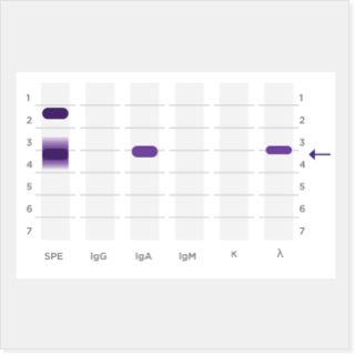
Different antibody classes of Igs in blood are identified using an IFE. If the presence of an M protein excess is identified by the Serum protein electrophoresis (SPEP) test, IFE will determine what subtype of M protein is present. Too much of 1 type of Ig is usually tied to specific types of blood cancer.

Serum free light chain assay
Igs are made up of smaller units called heavy chains and light chains, which bind together to form whole Igs. The presence of an M protein is consistent with the excess production of 1 type of light chain (kappa or lambda), which is detectable in the blood as a free light chain.
Imaging and biopsy testing for multiple myeloma

X-ray/bone survey
The inside of your body is able to be seen using X-ray imaging. The images show the parts of your body in different shades of black and white. Multiple myeloma will cause decreased bone density and appear as “punched-out” bone lesions. X-rays can help monitor disease.

Magnetic resonance imaging (MRI)
Organs and structures inside your body can be imaged using MRI. This technology uses strong magnets and radio waves to create images.

PET scan
PET scan can reveal the locations of cancer cells in different parts of the body using radioactivity. Radioactive glucose is put into your veins, which will be absorbed by cancer cells. Then, a special camera can detect the locations and activity of the cells.
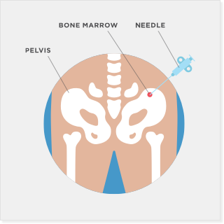
Bone marrow biopsy
A bone marrow biopsy may be used to confirm the presence and number of myeloma cells. This test can also be used to detect other blood-related diseases. People with multiple myeloma have too many plasma cells in their bone marrow.
Lab test value ranges
Reference ranges—values that are considered normal in healthy individuals—for common multiple myeloma lab tests are provided in the table below as a guide. Note that ranges may vary among laboratories.

Takeda Oncology
Here2Assist® patient support
We’re here for you throughout your treatment.
When multiple myeloma returns
Learn about signs and symptoms when multiple myeloma comes back